


 膜杰作
膜杰作 Star Staining


描述/背景(Background)
Programmed death ligand 1 (PD-L1) is the principal ligand of programmed death 1 (PD-1), a coinhibitory receptor that can be constitutively expressed or induced in myeloid, lymphoid, normal epithelial cells and in cancer. A key immune checkpoint is triggered when PD-1 (programmed cell death protein 1) engages its ligand PD-L1. As a result of this interaction, T cell activation is attenuated and an active immune response is prevented.
This mechanism is often co-opted by tumors. PD-L1 is upregulated in several tumor types and contributes to the malignancy of these cancers by interacting with PD-1 and inhibiting T cell activation. In this way, the tumors avoid detection and destruction by the immune system. Accordingly, PD-1 and PD-L1 have garnered much attention for their roles in tumor immunology and as immune-based therapeutic targets.
种属(Host Species)
Mouse
克隆号(Clone)
1G1
适用方法(Application)
IHC
效价(Property)
1:500
状态(State)
Liquid
阳性对照(Positive Control)
Human Tonsil Tissues
克隆类型(Clonality)
Monoclonal
分子别名(Synonym)
PDL1, PD-L1, CD274, B7-H1, B7H1
研究领域(Research Field)
Cancer Drug Targets
抗体来源(Source)
Mouse
同种型(Isotype)
IgG
储存(Storage)
Shipped at 4°C. Store at +4°C short term (1-2 weeks). Upon delivery aliquot. Store at -20°C long term. Avoid freeze / thaw cycle.
典型数据(Typical Data)
质控样本(Control Sample)
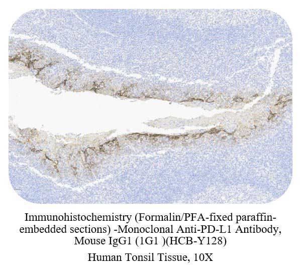
Immunohistochemical analysis of paraffin-embedded human tonsil tissue labeling PD-L1 with HCB-Y128 at 1/500 dilution, followed by Goat Anti-Rabbit IgG H&L (HRP) ready to use. epithelial portion of tonsillar crypts is observed as strong positive staining, follicular macrophages in the germinal center are observed as weak to medium positive staining, endothelial cells, fibroblasts, and surface epithelial cells are all observed as negative staining. Counter stained with Hematoxylin. Perform heat mediated antigen retrieval with Tris/EDTA buffer pH 9.0 before commencing with IHC staining protocol.
肿瘤样本(Cancer Sample)
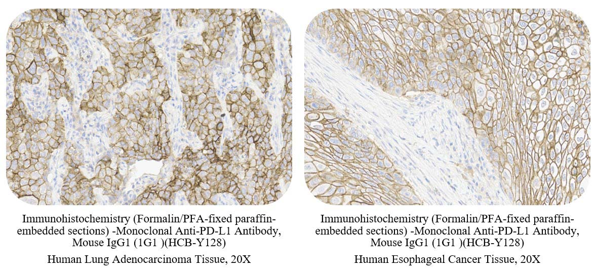
Immunohistochemical analysis of paraffin-embedded human cancer tissue labeling PD-L1 with HCB-Y128 at 1/500 dilution, followed by Goat Anti-Rabbit IgG H&L (HRP) ready to use. Membranous staining on human cancer tumor cells is observed. Counter stained with Hematoxylin. Perform heat mediated antigen retrieval with Tris/EDTA buffer pH 9.0 before commencing with IHC staining protocol.